
we constantly strive to improve
Our Research
Welcome to Our Research Page, where we showcase our ongoing commitment to pioneering advancements in Photodynamic Therapy (PDT) and its applications in the fight against cancer.
Minimally invasive and highly effective
Head and Neck PDT Clinical Summary
We present three compelling cases exemplifying the effective utilization of Photodynamic Therapy (PDT) in combating various forms of cancer, showcasing its versatility and potential impact on patient outcomes.
Over 1,500 patients treated with PDT
Over 1,500 patients (a mixture of presentations including primary, recurrent, and metastatic lesions )have been treated with PDT using Photofrin, HPD, ALA, or Foscan for head and neck cancers. The predominant histology is squamous cell carcinoma, but other histologies treated include mucosal melanoma, Kaposi’s sarcoma, adenocarcinoma, metastatic breast carcinoma, and adenoid cystic carcinoma.
Phase II Clinical Trials with Proven Efficacy
Several multi-institutional phase II clinical trials evaluating PDT treatment of head and neck cancers have demonstrated the efficacy of this minimally invasive therapy in the treatment of early oropharyngeal primary and recurrent cancers as well as the palliative treatment of refractory head and neck cancers.
Complete Clinical Response after One PDT Treatment
Of 518 patients treated with Cis, T1, or T2 cancers of the oral cavity, larynx, pharynx, and nasopharynx, 462 (89.1%) obtained a complete clinical response after one PDT treatment. Laryngeal cancers, comprising 171 patients in this group, obtained a durable complete response rate of 89% with up to a 16-year follow-up.
PDT is as effective as conventional therapies for the treatment of early (Cis, T1, T2) squamous cell cancers of the head and neck.
It is a promising therapy to be used in association with surgery to increase tumor-free margins and therefore increase cure rates.
Patient Case I
A diffuse oral cavity carcinoma in situ and early invasive squamous cell carcinoma.
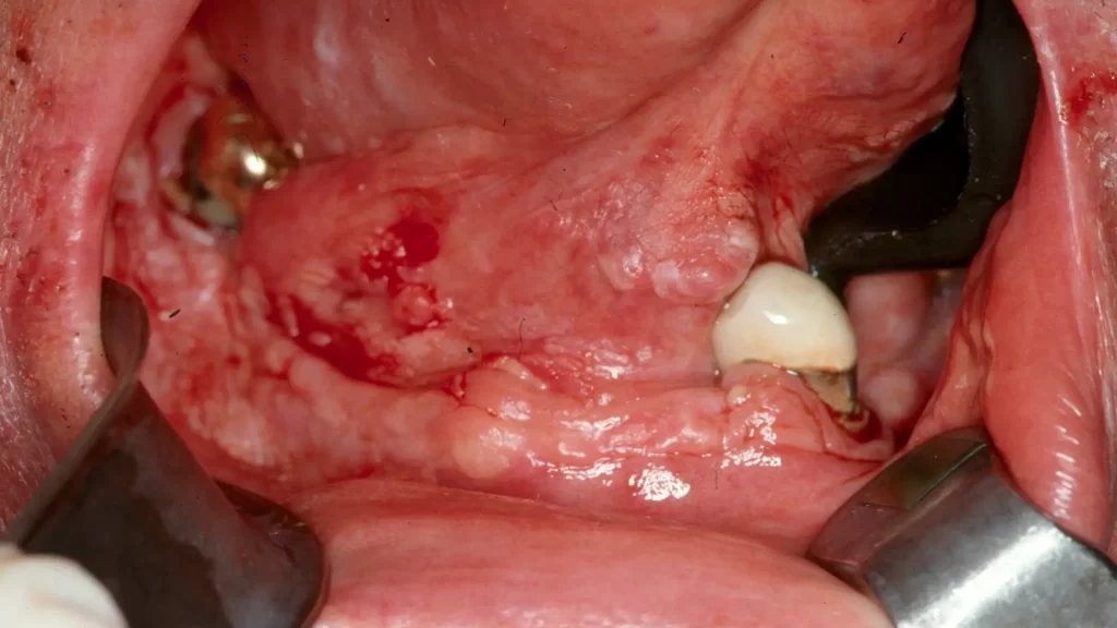
Picture 1
The tumor area involves the anterior and right lateral floor of mouth and lateral tongue extending onto the inner lip.
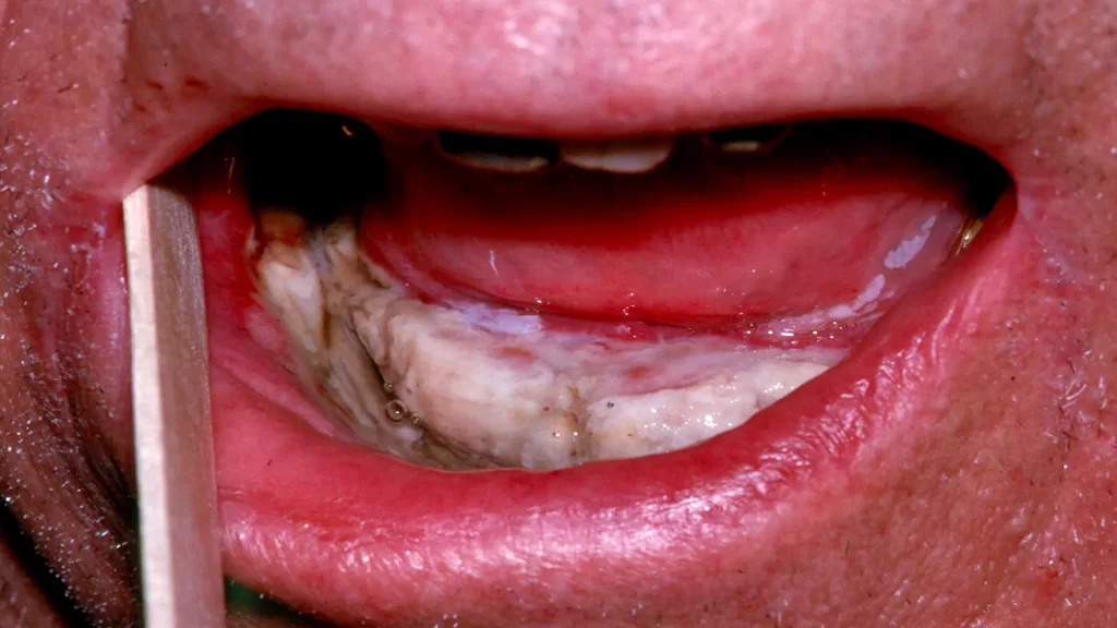
Picture 2
One week post PDT. The third picture is healed normal mucosa 6 weeks post PDT.
Picture 3
Healed normal mucosa 6 weeks post PDT.
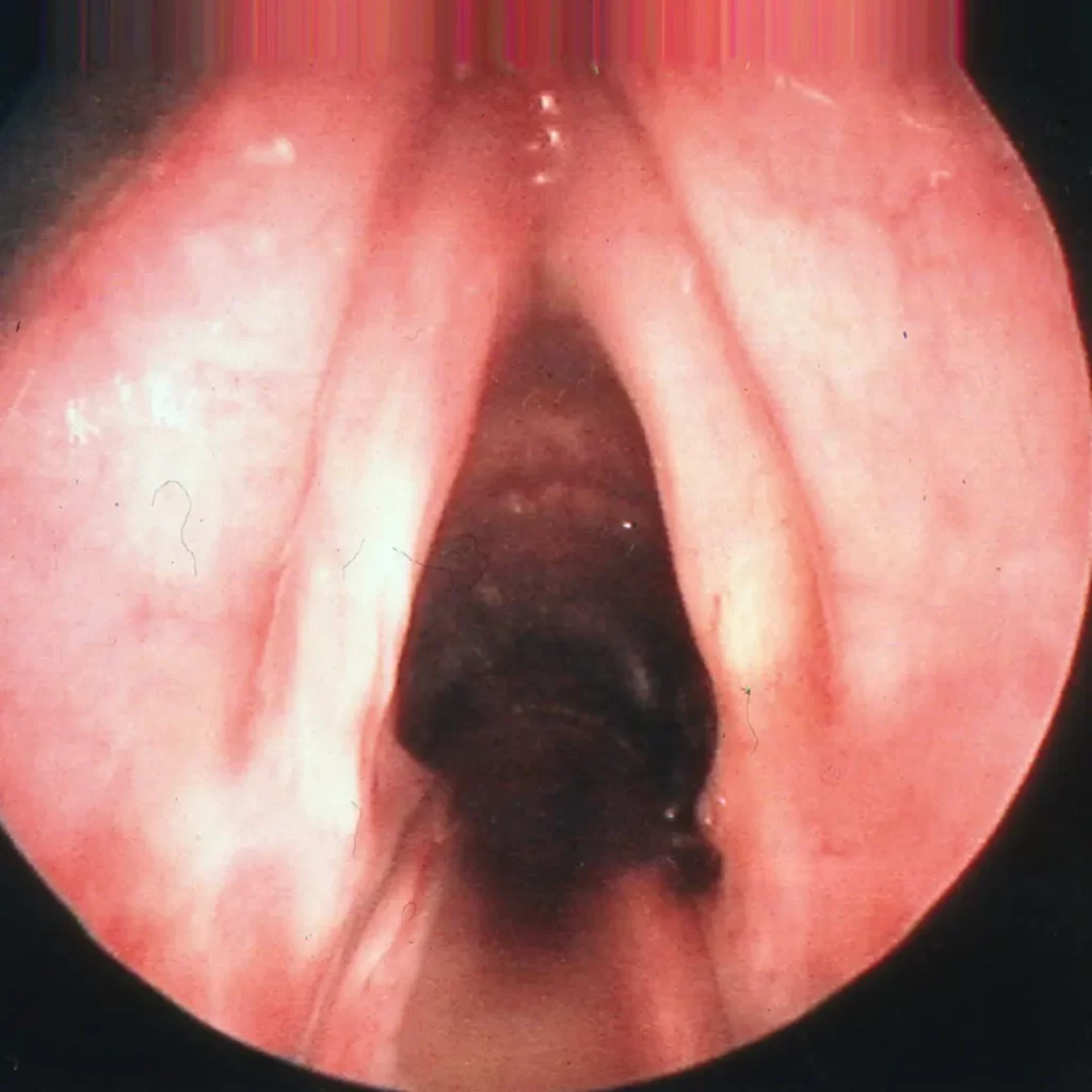
Patient Case II
A recurrent cancer after previous radiation.
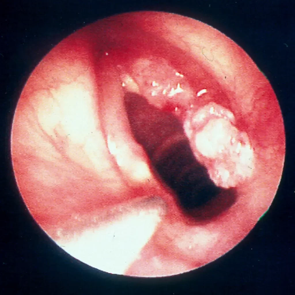
Picture 1
Recurrent right vocal cord cancer after previous radiation.
Picture 2
Six weeks post PDT with normal vocal cords.
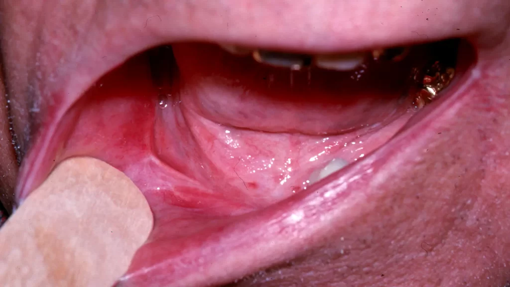
Patient Case III
A patient failed previous surgery, radiation, chemotherapy and surgery again.
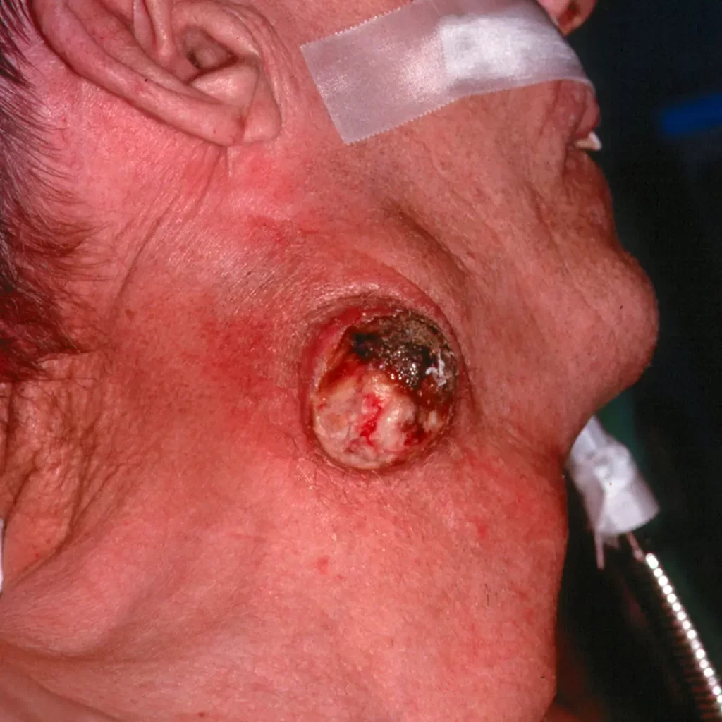
Picture 1
A patient with recurrent squamous cell cancer metastatic to the neck.
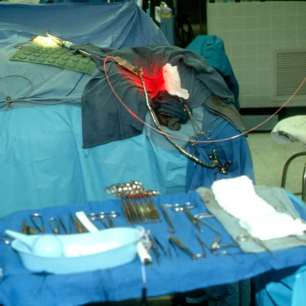
Picture 2
He was treated with a surgical resection and the surgical bed was treated with PDT to destroy any residual tumor.
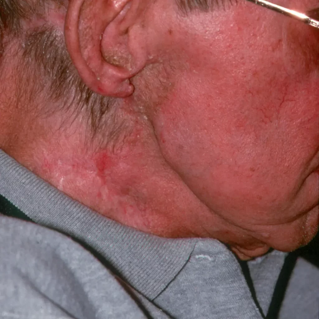
Picture 3
Healed normal mucosa 6 weeks post PDT.